Share this information on social networks
تبادل المعلومة
عبر المواقع الإجتماعية


Mobilization of primary metabolism
-
Enzymes lytiques: chitinase, glucanase. Faire la transgénèse avec les gènes de
ses enzymes. Des gènes codant pour ses protéines
et provenant de plantes (haricot, orge, luzerne) ont été
introduits chez le tabac. Néanmoins, les résistances
obtenues par cette voie sont partielles.
Pathogenesis-Related
(PR) Proteins
PR
proteins accumulate locally in the infected and surrounding
tissues, and also in remote uninfected tissues. Production
of PR proteins in the uninfected parts of plants can prevent
the affected plants from further infection. PR protein in
the plants was first discovered and reported in tobacco plants
infected by tobacco mosaic virus.
Later, these proteins were found in many plants. Most PR proteins in the plant species are acid-soluble, low molecular weight, and protease-resistant proteins. PR proteins depending on their isoelectric points may be
acidic or basic proteins but they have similar functions.
Most acidic PR proteins are located in the intercellular spaces,
whereas, basic PR proteins are predominantly located in the vacuole.
Currently PR-proteins were categorized into 17 families according to their properties and functions, including ß-1,3-glucanases, chitinases, thaumatin-like proteins, peroxidases,
ribosome-inactivating proteins,
defenses, thionins, nonspecific lipid transfer proteins, oxalate oxidase,
and oxalate-oxidase-like proteins.
Among these PR proteins chitinases
and ß-1,3-glucanases are
two important hydrolytic enzymes that are abundant in many plant
species after infection by different type of pathogens. The amount
of them significantly increase and play main role of defense reaction
against fungal pathogen by degrading cell wall, because chitin and
ß-1,3-glucan is also a major structural component of the cell
walls of many pathogenic fungi. ß-1,3-glucanases
appear to be coordinately expressed along with chitinases after
fungal infection. This co-induction of the two
hydrolytic enzymes has been described in many plant species, including
pea, bean, tomato, tobacco, maize, soybean, potato, and wheat.
Chitinase
induction
The fungal cell wall contains a cell membrane , which have a protective layer of chitin.
Abiotic agentssuch as chitosan, ethylene, ozone, wounding, polysaccharides, salicylic acid, salt solution, and UV light can induce higher expression of chitinases in plants.
Biotic agents
such as bacteria, insects, fungi, viruses, and fungal cell wall fragments can also induce the expression of chitinases in plants.
Studies of chitinases from bean, cucumber, pea, potato, sugar beet,
tomato, and tobacco have shown that the expression of chitinases is induced dramatically after infections.
The fungal cell wall contains a cell membrane with different proteins, a protective layer of chitin as well as glucan (mostly beta) and mannoproteins on its surface. Different fungal cell walls contain different glucans. The cell wall of Aspergillus fumigatus has beta-1,3- and beta-1,4-glucan and alpha-1,3-glucan. Candida albicans contains beta-1,3- and beta-1,6-glucan.
Induction usually occurs strongly at the point of infection and drops rapidly as the distance from the infection site increases.
The induction of chitinase can spread to adjacent tissues, resulting in a systemic acquired resistance that may enable the plant to protect itself from secondary infection.
CHITINASE AS PR PROTEIN AGAINST PATHOGENS. CASE OF MUSKMELON (MELON, البطيخ، الشمام) CUCURBITACEAE FAMILY
Plant
peroxidases and their involvement in resistance against fungi

Links:
Biotech-ecolo. net. SUPPORTS
CHAINE YOUTUBE Chaine Youtube (abonnement)
Plusieurs vidéos multilingues (+ s/titres) aux sujets des Biotechnologies et Biochimie
OUVRAGES
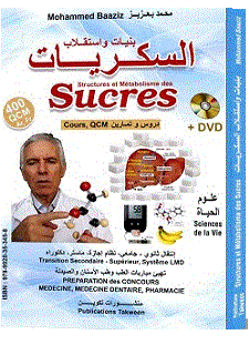
400 QCM et Concours (Ar, Fr) sur Structure et Métabolisme des
Sucres, M. Baaziz, 2018:
- Présentation
du livre
- Affiche du Livre
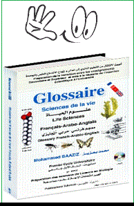
 Glossaire trilingues
Français-Arabe-Anglais
Glossaire trilingues
Français-Arabe-Anglais
- Livre 'Sciences de la vie. Protéines et Enzymes' (+ DVD),Baaziz,
2013: QCM corrigés, Exercices corrigés, Contrôles
corrigés en Biologie cellulaire et Biochimie pour S1, S3, S4
et S5.
Biotechnologies.QCMs, Examens ..

biotech-ecolo. ANNONCES
ENSEIGNEMENT DE MASTER
-
- Cours du Master Biotechnologies et Amélioration des Plantes,
Marrakech
-
Travaux pratiques Master Biotechnologies et Amélioration des Plantes, Marrakech
-
Master course 'Plant resistance against pathogens'
COURS, WORKSHOPS
- Annonces de Cours
- Annonces de Workshops
BOURSE, POSTDOCS
BOURSES
POSTDOCS
NEWSLETTER
NEWSLETTER
BIOTECHNOLOGIES
NEWSLETTER
BIOCHIMIE


